Our Technology
“An Eye Exam Is More Than Just Your Vision”
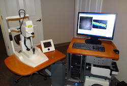
Union City Eye Care Diagnostic Instrumentation
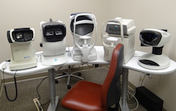 Preliminary Testing
Before you see the doctor for your eye exam, a thorough history and pertinent data is obtained. Technicians use the most modern and precise instrumentation available to evaluate your vision and eye health including intraocular pressure, color vision, binocular vision, corneal curvature, static refractive state, wavefront aberrometry, peripheral vision screening visual fields, and ultra wide field retinal imaging. Some of the instrumentation used in this process includes: Marco OPD III (corneal/refractive) Wavefront analyzer, Stereo Optical vision screener, Canon non-contact tonometer, Zeiss FDT visual fields, and Optos optomap.
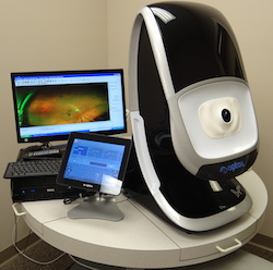 Optos optomap Ultra Wide Field Retinal Imaging
Optomap is one of the newest modalities for imaging the posterior section of the eye, allowing up to a 200 degree field of view and observation of the retina, optic nerve head, and blood vessels. It is beneficial in detecting and monitoring changes in pathologies of the eye including macular degeneration, retinal disease, diabetic retinopathy, vascular changes, and much, much more.
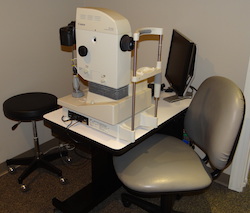
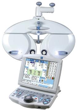 Marco Total Digital Refraction System
This innovative refraction system is a state-of-the-art automatic refractor that helps us to better evaluate your unique visual needs and achieve the most accurate prescriptions in a fraction of the time. We can perform additional tests to compare your old and new prescriptions and you will notice that it is easier for you to tell us “Which is better—1 or 2?”
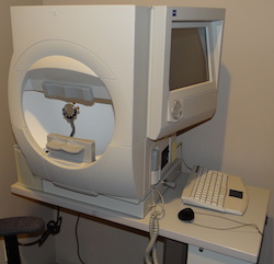 Zeiss Humphrey Visual Fields (VF)
Visual fields is a neurologic function test of the visual pathway between the eye and the brain useful in detecting and monitoring glaucoma, multiple sclerosis, optic nerve disease, trauma, and tumors that involve the visual system.
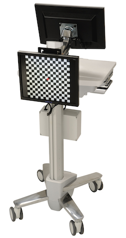 Diopsys Visual Evoked Potential (VEP)
![]()
Visual evoked potential is an objective neurologic test to detect or rule out optic nerve and visual pathway pathologies such as glaucoma, multiple sclerosis, ischemic optic nerve neuropathy, and more.
Images provided by Diopsys, Inc.
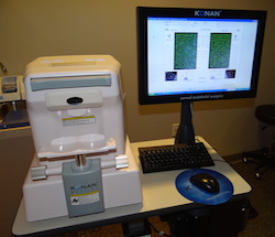 Konan Specular Microscopy with Cell Count (SMCC)
Specular microscopy examines the back layer of the cornea called the endothelium and actually counts the cells. Since we lose cells continuously and they are never replaced, some persons will develop corneal disease due to excessive cell loss. With this instrument, the progression of cell loss can be monitored and treatment recommended if needed.
 Retinal Photography
Fundus (retinal) Imaging
Retinal photography is used to image the back of the eye including the retina, the front of the optic nerve, and the blood vessels. It is also used to document lesions or conditions such as diabetic retinopathy, optic nerve atrophy, nevi, macular degeneration, etc.
Stereo Disk Imaging
Stereo disk imaging provides a 3 dimensional image of the front of the optic nerve which is made up of hundreds of thousands of individual nerve fibers and is the beginning of the visual pathway. Conditions that can damage the optic nerve include glaucoma, optic nerve atrophy, MS, optic nerve drusen, and many others.
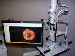 External (anterior segment) Photography
External photography uses a biomicroscope and a digital camera to image specific structures in the front of the eye such as the cornea, conjunctiva, iris, and more.
 Heidelberg Engineering Spectralis Ocular Coherence Tomography (OCT)
OCT of Optic Nerve, Retina, or Anterior Segment
With OCT, the layers of the retina, nerve fibers that make up the optic nerve, and the structures in the front of the eye can be imaged using a scanning laser and computer assisted tomography. Both 2 and 3 dimensional images, analogous to a CT scan, are obtained. OCT aids in the diagnosis and management of such conditions as glaucoma, macular degeneration, diabetic and hypertensive retinopathy, tumors of the eye, retinal holes and tears, and numerous other eye health and vision conditions.
Auto-Fluorescence
Auto-fluorescence is a study of the function of retinal pigment epithelial cells and aids in the detection and monitoring of macular degeneration and other pathologies involving the outer retinal layers.
–At Union City Eye Care, the following tests are also available.
Tonometry
Tonometry measures the pressure in the eye. High eye pressure can damage the optic nerve leading to the most common form of glaucoma.
Serial Tonometry
This test allows us to take measurements to determine if the eye pressure may be spiking at different times of the day or night, which would indicate inadequate control of the intraocular pressure and require a change in therapy.
Pachymetry
Pachymetry measures the thickness of the cornea which is important in evaluating its effect on eye pressure and determining the presence of corneal edema related to trauma, infection, or contact lens wear.
Gonioscopy
Gonioscopy allows the evaluation of specific structures inside the eye that control intraocular pressure or may be damaged or degenerate with age.
Extended Ophthalmoscopy
Dilation for extended ophthalmoscopy allows us to view as many structures in the back of the eye as possible, such as the retina, front of the optic nerve, vitreous, and retinal blood vessels. An annual dilated retinal exam is recommended for everyone to better evaluate eye health. It is sometimes recommended more often in cases of risk due to health issues or previous findings.
Corneal Topography (CT)
Corneal topography is mapping the shape of the front of the eye looking for any irregularities or unusual shape of the corneal surface. This is helpful in detecting conditions such as keratoconus or other degenerations of the cornea.
Ophthalmodynamometry (ODM)
ODM allows the evaluation of blood flow and circulation to the eye.
Sensorimotor Exam (SMEX/EOM)
SMEX tests muscle function of each eye, looking for any weakness or limitations in any of the twelve extraocular muscles or the nerves that control them.
B Scan Ultrasound
B Scan uses sound waves (ultrasound) to image structures inside the eye including the retina, choroid, vitreous, and ciliary body.
Tear Lab Osmolarity Test
Tear Lab Osmolarity Test is an objective and quantitative measurement of the osmolarity of human tears to aid in the diagnosis of dry eye disease.